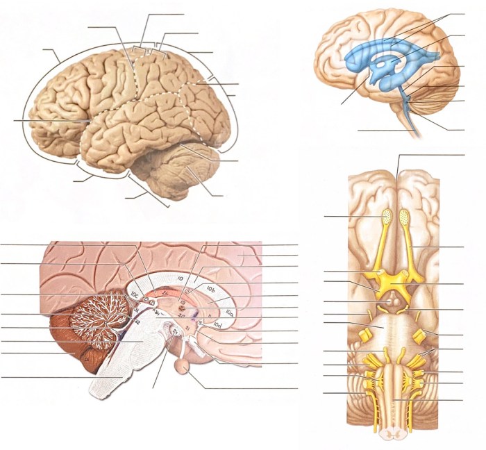
Exercise 17 gross anatomy of the brain and cranial nerves – Exercise 17: Gross Anatomy of the Brain and Cranial Nerves embarks on a fascinating journey into the intricate world of neuroanatomy, providing a comprehensive examination of the brain and its associated cranial nerves. This exploration delves into the structural and functional aspects of these vital components, offering a profound understanding of their significance in human physiology and clinical practice.
Through a meticulous analysis of external and internal brain features, we unravel the complexities of the cerebrum, cerebellum, and brainstem, gaining insights into their specialized roles. Furthermore, a thorough examination of the 12 pairs of cranial nerves reveals their diverse sensory, motor, and autonomic functions, emphasizing their critical contributions to bodily processes.
1. Introduction to Gross Anatomy of the Brain and Cranial Nerves
The study of the gross anatomy of the brain and cranial nerves is essential for understanding the structure and function of the nervous system. The brain is the central processing unit of the body, responsible for controlling movement, thought, emotion, and memory.
The cranial nerves are responsible for transmitting sensory and motor information between the brain and the rest of the body.
The brain is divided into three main regions: the cerebrum, the cerebellum, and the brainstem. The cerebrum is the largest part of the brain and is responsible for higher-order functions such as cognition, language, and memory. The cerebellum is responsible for coordination and balance.
The brainstem is responsible for vital functions such as breathing, heart rate, and blood pressure.
There are 12 pairs of cranial nerves. They are classified into three groups: sensory nerves, motor nerves, and mixed nerves. Sensory nerves transmit sensory information from the body to the brain. Motor nerves transmit motor commands from the brain to the muscles.
Mixed nerves transmit both sensory and motor information.
2. Methods and Techniques for Studying Gross Anatomy

The traditional method for studying the gross anatomy of the brain and cranial nerves is dissection. Dissection involves carefully removing the skin and other tissues to expose the underlying structures. Dissection is a valuable technique for learning about the anatomy of the brain and cranial nerves, but it can be time-consuming and destructive.
Modern imaging techniques, such as MRI and CT scans, provide a non-invasive way to study the gross anatomy of the brain and cranial nerves. MRI scans use magnetic fields and radio waves to create detailed images of the brain. CT scans use X-rays to create detailed images of the brain.
MRI and CT scans are valuable techniques for diagnosing and treating neurological disorders.
3. External and Internal Features of the Brain: Exercise 17 Gross Anatomy Of The Brain And Cranial Nerves

The external features of the brain include the cerebrum, the cerebellum, and the brainstem. The cerebrum is the largest part of the brain and is divided into two hemispheres by the longitudinal fissure. The cerebellum is located at the back of the brain and is responsible for coordination and balance.
The brainstem is located at the base of the brain and is responsible for vital functions such as breathing, heart rate, and blood pressure.
The internal features of the brain include the ventricles, the white matter, and the gray matter. The ventricles are four fluid-filled cavities that are located within the brain. The white matter is composed of myelinated nerve fibers that connect different parts of the brain.
The gray matter is composed of cell bodies and dendrites.
4. Structure and Function of Cranial Nerves

There are 12 pairs of cranial nerves. They are classified into three groups: sensory nerves, motor nerves, and mixed nerves. Sensory nerves transmit sensory information from the body to the brain. Motor nerves transmit motor commands from the brain to the muscles.
Mixed nerves transmit both sensory and motor information.
The cranial nerves are responsible for a wide range of functions, including vision, hearing, smell, taste, touch, and movement. They also control vital functions such as breathing, heart rate, and blood pressure.
5. Anatomical Relationships and Clinical Correlations

The brain and cranial nerves are closely related anatomically. The cranial nerves pass through the skull and connect to the brain at the brainstem. The anatomical relationships between the brain and cranial nerves are important for understanding how the nervous system functions.
Understanding the gross anatomy of the brain and cranial nerves is essential for diagnosing and treating neurological disorders. For example, a stroke can damage the brain and cause neurological symptoms such as paralysis or speech problems. A tumor can compress the cranial nerves and cause symptoms such as vision loss or hearing loss.
Essential Questionnaire
What is the significance of studying the gross anatomy of the brain and cranial nerves?
Understanding the gross anatomy of the brain and cranial nerves provides a foundation for comprehending their functions, interconnections, and clinical implications in neurological disorders.
How many pairs of cranial nerves are there, and what are their classifications?
There are 12 pairs of cranial nerves, classified into sensory, motor, and mixed nerves based on their primary functions.
What are the advantages of using modern imaging techniques in neuroanatomy?
Modern imaging techniques, such as MRI and CT scans, offer non-invasive visualization of brain structures, allowing for detailed examination and diagnosis of neurological conditions.